

By Elisabeth Lidwien, J.M.M. Verdegaal, Gordon S. Howarth, Todd J. McWhorter, Catherine J.G. Delesalle
Graphics: The authors
Heat stress is an important performance and welfare issue for exercising horses. The process of thermoregulation is crucial for equine athletes. Excessive metabolic heat generation during exercise, combined with inefficient heat dissipation, can lead to hyperthermia if not detected in time and not effectively managed.
Heat stress is an important performance and welfare issue for exercising horses. The process of thermoregulation is crucial for equine athletes. Excessive metabolic heat generation during exercise, combined with inefficient heat dissipation, can lead to hyperthermia if not detected in time and not effectively managed. Accurately monitoring heat generation during exercise allows for early preventative intervention as body temperature rises. Skin temperature monitoring is commonly used as a non-invasive method to assess body temperature responses pre- and post-exercise.
To date, few studies have used infrared thermographic techniques to monitor body temperature continuously during exercise under laboratory conditions and in the field. In reviewing these results, the accuracy of measuring skin temperature as a reliable indication of overall body temperature is discussed. This commentary summarizes the results of studies measuring surface skin temperature in horses, particularly using infrared thermography for exercise-focused monitoring in the field.
Abstract
Hyperthermia and exertional heat illness (EHI) are performance and welfare issues for all exercising horses. Monitoring the thermoregulatory response allows for early recognition of metabolic heat accumulation during exercise and the possibility of taking prompt and effective preventative measures to avoid a further increase in core body temperature (Tc) leading to hyperthermia. Skin temperature (Tsk) monitoring is most used as a non-invasive tool to assess the thermoregulatory response pre- and post-exercise, particularly employing infrared thermographic equipment. However, only a few studies have used thermography to monitor skin temperature continuously during exercise. This commentary provides an overview of studies investigating surface skin temperature mainly by infrared thermography (IRT) during exercise.
The scientific evidence, including methodologies, applications, and challenges associated with (continuous) skin temperature monitoring in horses during field exercise, is discussed. The commentary highlights that, while monitoring Tsk is straightforward, continuous Tsk alone does not always reliably estimate Tc evolvement during field exercise. In addition, inter-individual differences in thermoregulation need to be recognized and accounted for to optimize individual wellbeing. With the ongoing development and application of advanced wearable monitoring technology, there may be future advances in equipment and modeling for timely intervention with horses at hyperthermic risk to improve their welfare. However, at this point, infrared thermographic assessment of Tsk should always be used in conjunction with other clinical assessments and veterinary examinations for a reliable monitoring of the welfare of the horse... To read the complete article you need to be a subscriber
CLICK HERE TO SUBSCRIBE TO BREEDING NEWS
SUBSCRIBERS CAN READ THE COMPLETE ARTICLE BY LOGGING IN AND RETURNING TO THIS PAGE
Field exercise is an integral aspect of equine sports and competition where horses face a myriad of physiological and environmental challenges. Efficient thermoregulation is paramount to ensure optimal performance and prevent heat-related stress in equine athletes. The incidence of heat stress and exertional heat illness (EHI) is projected to rise further due to the ongoing effects of global climate change. Climate change is a generally accepted phenomenon, and the scientific community is warning the world about its long-term consequences both for human and animal health [1,2,3]. Particularly, more acute changes in environmental conditions such as heat waves, entail that athletes may need to perform under circumstances to which they are not acclimatized. These sudden increased ambient temperatures (Ta) are the dominant external risk factor for heat stress for human and equine athletes [1,2,3].
EHI is associated with various kinds of exercise and impacts the overall well-being of all mammals, including humans, and may lead to exertional heat stroke (EHS) and ultimately death. In exercising human athletes, EHS is among the top three causes of sudden death, and in summer, it is the number one cause of athlete death in the USA [4]. For example, EHI is common in American football, with an average incidence rate ranging from 0.01 to 0.4%. Heat monitoring studies being developed to investigate the EHI problem in human athletes have used many monitoring approaches such as ingestible thermo-monitoring pills or a rectal thermistor, skin temperature (Tsk) sensors, and measures of subjective perception of heat load [5].
Both EHI and EHS are also problematic conditions in equine athletes and can occur during a wide variety of different types of exercise [6]. However, heat exhaustion triggered by thermoregulatory-induced physiological feedback failure is most commonly reported when horses need to perform submaximal exercise during extended time intervals such as endurance rides. The prevalence of metabolic disorders in endurance horses ranges from 2% to 15% [7,8,9,10,11,12,13,14].
In comparison, for maximal intensity exercise, EHI screening studies have recently reported a prevalence between 0.1% and 1.1% in racehorses in Australia, the UK, and Japan [15,16,17,18,19]. A study in eastern Australia focused on selected EHI cases post-exercise at the racetrack and suggested an EHI incidence of up to 9.5% during hot summer months [17]. The latter study used four severity levels of EHI (1 = least severe) reported by Brownlow et al. [20] and concluded that 96% of horses could be categorized as level 1. This suggests that many EHI cases of low-level severity with vague clinical signs may have been overlooked in the past. At present, the association between EHI and exercise injuries leading to fatalities is not well understood, and hence the roles that heat exhaustion and EHI play are unknown.
All equestrian sports are facing growing scrutiny from both the general public and animal welfare organizations. When unfortunate incidents result in fatalities, spectators are swift to criticize sporting and racing events and increasingly call for safeguarding the well-being of sports horses in order to maintain the ‘social license’ to operate in equestrian sports [21]. Consequently, from an animal welfare perspective, it is paramount and timely to prevent occurrence of heat stress on all occasions. The aforementioned welfare issues are a concerning reality that is motivating research groups worldwide to develop practical methods for reliable monitoring during field exercise to ensure the thermoregulatory welfare of horses [21,22,23,24,25,26]. To achieve this, there is a need for minimally invasive body temperature monitoring systems, also termed ‘heat monitoring’. The ultimate goal of monitoring is to accurately detect a potentially dangerous rise in core body heat in real-time such that effective preventative actions can then be taken.
In an ideal world, all temperature monitoring equipment including wearable technological tools should be non-invasive and enable horse owners and veterinarians to have a continuous real-time view of the evolution of body temperature during exercise in the field [22,23,24,25]. An approach that is gaining popularity for monitoring the thermoregulatory response is the follow-up of the Tsk in horses performing in the field exercise using wearable Tsk equipment [24]. While this equipment provides output of physiological data, the question remains whether these data have physiological relevance for the evolution of the core body temperature, which is the core point of interest to safeguard welfare in a reliable way. This article reviews and comments on the benefits and challenges of monitoring thermoregulation in exercising horses using non-invasive surface Tsk, particularly through infrared thermography.
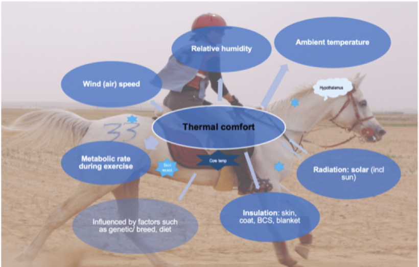
Factors affecting thermoregulation in horses and its feedback control mechanisms including thermoreceptors in the skin (spread over body, examples displayed as bright-blue 6-point stars), and the core temperature receptors (dark-blue 6-point star) which provide information to the hypothalamus (displayed as light-blue call-out), which adjusts the responses either to produce or to lose heat to ensure thermal comfort; BCS: body condition score.
2. Thermoregulation: core vs shell temperature
Thermoregulation is the internal physiological process which balances metabolic heat load and the exchange of this heat with the environment. Overall body temperature in mammals is divided into an inner core body temperature (Tc) and an outer shell temperature. Tc in horses is regulated within a narrow range (37.4–38.0°C), while shell temperature varies more widely in response to ongoing central thermoregulatory processes [24,26]. The inner core temperature refers to deep-body temperatures, while shell temperature includes intramuscular, subcutaneous, and surface skin temperatures (Tsk). Heat is moved between core and shell areas to equilibrate heat production and heat loss and thus regulate the overall body temperature. Consequently, temperature is not uniform at different locations in and on the body.
Thermoregulatory processes are controlled via neurophysiologic control centers in the hypothalamus. Both peripheral and central thermoreceptors are nerve endings that respond to changes in Tc and Ta, all in communication with the hypothalamus (Figure 1). The peripheral thermoreceptors in the skin detect the peripheral shell temperature and respond to a range of external temperatures from cold to hot (5–60°C), with cold thermoreceptors being the most numerous.
3. Mechanism of metabolic heat loss
During exercise, heat originating from exercising muscle groups will be dissipated to the surrounding environment through different tissues and by using transportation via the bloodstream towards the skin surface. H will then be released into the surrounding environment through four mechanisms: radiation, conduction, convection, and evaporation (Figure 1). Therefore, the surface area (cm2) of the body is a critical factor in determining rates of heat exchange.
Because heat exchange with the environment is proportional to the relative size of the body surface area (BSA), the rate of heat exchange per unit of body mass (BM) is largest in the smallest animal (if other variables are equal). For example, the BSA-to-volume ratio is 50% less in a 500 kg horse when compared to humans [26,27]. This lower BSA-to-BM ratio of horses results in greater demands being imposed on their thermoregulatory system during exercise [28,29,30]. Only if Tsk is higher than the surroundings can the body lose heat by radiation, conduction, and evaporation.
Among these heat-loss pathways, the most critical and pivotal for horses is the activation of sweat glands to evaporate sweat from the skin surface [27,29,31,32,33,34]. The efficacy of heat dissipation via this evaporative process relies on the thermal contrast between locally perfusion-based Tsk and its immediate surroundings, with consideration of variables such as vapor pressure, airflow, and solar radiation, especially during outdoor exercise [17,28,35] (Figure 1). Consequently, when considering using Tsk as a method for monitoring temperature, it is essential to anticipate that all pathways for dissipating H to the environment can influence the Tsk data output.
Additionally, horses are hindgut fermenters, which entails that important fermentation processes take place within the gastro-intestinal (GI) system [26]. These processes are recognized for their propensity to produce an enormous amount of heat which in turn represents an additional challenge for the thermoregulatory system. It is quite plausible that the type of dietary load (composition of intestinal content) inside the GI system of a horse performing exercise has its impact on Tc evolution at that time point [26]. How quickly the latter is translated towards the outer shell temperature is currently unknown, but again, requires notice as a factor that may influence the Tsk data output. From a comparative perspective, in humans it is known that food containing high amounts of fats, complex carbohydrates, and proteins stimulate the Tc increase during digestion [26].
4. Field-based thermoregulation research – non-invasive monitoring
Until recently, little was known about how thermoregulation functions in real-life circumstances during field exercise and recovery in different equine sports disciplines. An important reason for this was the complete lack of effective, reliable temperature monitoring equipment that can be safely and comfortably applied during field exercise.
In the past, most equine thermoregulatory research has been performed either under laboratory conditions, for example on a treadmill [36,37,38], or under field conditions, only focussed on the pre- and post-exercise period. A few studies have aimed at continuous monitoring of the Tc during exercise and recovery in real-time field training and competitions. One of those studies used intra-uterine temperature loggers as a nonsurgical, minimally invasive method [39], an approach which only allows for monitoring in female horses. Recently, a reliable novel method is the oral administration of a small telemetric GI temperature pill to monitor Tc on a continuous basis [40,41,42,43].
The aforementioned field studies reveal that in-the-field exercise monitoring is essential to obtain a holistic view on how the thermoregulatory system actually works. This is not only challenged by the performed exercise itself, but also by the environmental conditions in which environmental temperature and humidity are pivotal factors [43]. Additionally, the terrain itself influences exercise intensity. For example, the presence of slopes and descents in a competition course as well as varying properties of the surface over the course, such as sand or mud, are challenging for optimal exercise capacity [43]. These factors must all be accounted for when monitoring Tc during exercise, and for that purpose, monitoring thermoregulation during field exercise has been shown to be more reliable than lab-based testing [43].
In addition to thermoregulation monitoring, it is important to consider thermal comfort when assessing the welfare of horses (Figure 1). Thermal comfort refers to a state in which horses can still successfully thermoregulate, but may already feel thermally uncomfortable. This is similar to evaluating a perception of heat exertion during heat monitoring in human athletes. Many factors that impact thermal comfort, such as solar radiation, skin insulation, and wind speed, can also affect Tsk values.
5. Skin temperature measurement
Non-invasive methods such as Tsk measurement are of significant interest and have found various applications. Generally, four types of temperature sensor equipment are employed in thermoregulation research: thermometers, thermistors, thermocouples, and infrared thermography (IRT) devices. Of these, IRT has received the most extensive attention in recent equine studies and is commonly used remotely (non-contact temperature sensor) [22,44,45,46,47,48,49,50,51,52,53,54,55,56].
Digital temperature sensors may include technology that comprises a logger consisting of a temperature sensor in a stainless-steel computer chip with an enclosed battery [57]. Thermistors are temperature-sensitive resistors that change resistance with temperature and have been used in several equine studies [34,58,59,60]. Both digital temperature sensors and thermistors have a small size, fast response time, and relatively low cost. However, they are invasive, are limited to point measurements, and may alter the local skin conditions.
Thermocouples are temperature sensors based on the Seebeck effect, where a voltage is generated in response to a temperature difference (thermo-electric sensors) and have been used in some equine studies [57,61,62]. In addition, fiber optic sensors can be used to record temperature by measuring the change in light transmission properties through an optical fiber to measure temperature [63].
In human thermoregulatory research, conductive textiles have received increasing attention recently. These are textiles with embedded conductive fibers that can be used to measure Tsk. However, this technique still encounters many calibration challenges, and the accuracy of data output is influenced by textile placement [63]. In addition, the dermal patch technology is a small adhesive patch with integrated sensors applied to the skin surface [63]. Lastly, IRT measures the infrared radiation emitted by an object to create a temperature map [22] and has multiple applications in equine medicine, including detecting inflammation and musculoskeletal injuries such as lameness, tendonitis, and laminitis [51,64,65]. In several studies, IRT has been used as an indicator of stress, but those studies investigated only a limited number of horses (five horses) [44,46,65,66] and one study investigated the correlation of increased Tsk with blood lactate concentration post exercise in 30 horses [67]. Lastly, infrared contact sensors (IRCSs) are contact-based sensors using infrared technology to measure Tsk [24,60].
6. Skin temperature at point(s)-in-time or continuous monitoring during rxercise
Changes in Tsk during field exercise are complex. As exercise continues, Tsk varies by body region and may be transient and rapidly changing in relation to local heat exchange from the muscles to the environment, an increased sweat rate, and forced convection (wind), all highly influenced by Ta.
6.1. Tsk measurement at a point-in-time (pre-exercise and post-exercise): The most common method to record Tc in field competition is serial measurement of the rectal temperature (Tre) pre- and, more often, only post-exercise. These are “snapshots” (points-in-time) of temperature evolvement. Importantly, research has shown that Tre lags behind or is always lower than Tc [28,42]; consequently, serial Tre measurement does not allow for early-stage remedial intervention. Importantly, there is also a clear lack of correlation between Tsk and Tc [24].
To date, several methods are employed for monitoring Tsk such as infrared thermography using cameras. While handheld IRT cameras are becoming increasingly popular in equine sports medicine due to their non-invasiveness, most exercise studies involve point-in-time Tsk measurements only post-exercise and without pre-exercise and intra-exercise periods [22,30,45,46,47,48,52,53,58,68,69,70]. A non-exhaustive overview of Tsk studies associated with exercising horses, either monitoring non-continuously or continuously in horses, is presented in Table 1.
A systematic review of 45 Tsk studies of human endurance athletes reported that only a few studies involved continuous temperature monitoring [23]. From a physiological viewpoint, it is more than plausible that not only the actual temperature increases, but also the rate of increase and the time profile which are important parameters to reliably estimate how an individual athlete copes with heat load. Those parameters remain a “black box” when only non-continuous temperature monitoring is applied. Some human studies have applied highly frequent monitoring intervals. Two studies monitored Tsk every 30 s, one study every 180 s, and four studies every 5 min [23]. These human studies highlighted essential inter-individual differences concerning the Tsk response time profile [23]. Similarly, significant inter-individual Tsk differences have been reported in all mammals [33], including a recent equine exercise study [24]. In addition, Tsk in 21 horses monitored at rest also revealed significant inter-individual differences, which entails that defining “thermo-comfort” reference ranges actually needs a customized approach [50].
In horses, several studies have revealed possible correlations between recorded point-in-time Tsk values and various other physiological parameters [24,45]. For example, a study involving eight endurance horses analyzed the association between endurance training intensity and Tsk evolvement using IRT cameras focused at different locations on the body. This revealed that Tsk at the coronary band increased with training intensity, but maximum Tsk did not [45].
Another study monitoring endurance horses revealed that a higher Tsk at the end-of-exercise period was associated with a lower Tc at the end-of-recovery period (60 min) [24]. In addition, there was no significant correlation between Tsk at the end of exercise and the duration of HR recovery to 60 bpm [24].
Several hypotheses could be considered to explain the marked difference between Tsk and Tc, both with respect to temperature values and evolvement over time. For example, it could be hypothesized that the raised Tsk functions as an ‘early whistle blower’ for the launch of an active thermoregulatory response to anticipate the increased Tc, and once the metabolic heat is successfully dissipated, the Tc decreases. This argument could be coupled with the effect of cooling-down measures being applied post-exercise, which may be more prominent when Tsk is greater and ultimately result in higher dissipation of metabolic heat and swifter reduction of Tc. However, the important fact that both temperature values and time evolvement patterns importantly differ between Tsk and Tc shows that the physiological link between both is much more complicated than initially anticipated in studies investigating the many monitoring wearables on the equine market.
Interestingly, an immediate post-race, high point-in-time IRT Tsk measurement >39°C in the neck region of racehorses exercising in a warm-hot Ta has been advocated as an early indicator for increased risk for development of post-exercise EHI [48]. Possibly in the case of the manifestation of a narrow temperature gradient between Tc and Tsk, the capacity of the equine body to transfer heat to the skin is greatly reduced and thus most probably hinders heat dissipation through evaporation [24,48]. Therefore, additional research has tested the effectiveness of different cooling-down techniques with the obvious goal of increasing the temperature gradient between the Tsk and the Tc post racing.
A recent study evaluated five cooling methods for racehorses – walking with and without air fans, intermittent application of cold water with or without scraping, and continuous pouring of tap water over the body [52]. The study revealed a mean Tsk of 40 °C at the end-of-race exercise in a warm environment (mean Ta 31.8°C) [52] and a Tsk lower than 34.5°C at the end of moderate intensity exercise in a moderate environment (mean Ta 24°C) [59]. Of considerable concern is the occurrence of a markedly increased Tsk post-exercise (>39°C) which could indicate the horse is potentially already manifesting mild to severe manifestations of EHI.
Significantly, all of the aforementioned studies were performed under a wide range of Ta, from hot to cool conditions, and tested large disparities in exercise intensity ranging from racing to endurance exercise [24,45,48]. The pattern of Tsk and its variability can partly be attributed to acute fluctuations in blood flow associated with differing exercise intensities [32,60].
In racehorses executing high-intensity, short-duration exercise, heat dissipation will typically occur post-exercise, while endurance horses manage heat dissipation throughout their submaximal prolonged exercise and their mandatory monitored rest periods [43,48,71,72].
6.2. Continuous Tsk monitoring during exercise: To date, study results have clearly shown that the relationship between Tsk and Tc is very complex [32,33,72], and those studies that involve continuous temperature monitoring are pivotal to better understand this complexity. Only eight studies have applied continuous Tsk monitoring during exercise, of which four studies involved a controlled laboratory environment using treadmills [36,37,38,44] (Table 1). These laboratory-based investigations cannot address the authentic environmental conditions encountered by sports horses during open-air competitions. By comparison, different important variables can be deduced from the analysis of the IRT data time profiles during exercise in the field such as the average, maximum and minimum Tsk, standard deviation, and delta Tsk (Tsk variation during the exercise) [24].
Currently, only a few studies have tried to correlate both continuous Tc (through either arterial, venous blood, or GI pill temperature) and Tsk in an attempt to analyze if Tsk could function as a proxy for the core body thermoregulatory response [24,36,37,38,62] (Table 1). Importantly, the four laboratory-based treadmill investigations consistently affirmed the absence of a significant correlation between Tsk and Tc. For instance, two submaximal exercise studies used arterial blood temperature as a reference Tc to compare the impact of different environmental conditions varying from cool, dry (room Ta and 45% relative humidity (RH)) to hot, humid (34°C, 85% RH) on the thoracic surface Tsk using thermocouples and revealed dissimilarities between Tsk and Tc [61,62].
Two high-intensity exercise studies investigated tail surface Tsk evolvement response to cooling-down techniques (6 times cold water shower for 30s) and acclimation (30°C and 80% RH over 15 days), respectively, and both demonstrated clear distinctions between Tsk and Tc recordings [37,38].
One other study did not use Tsk monitoring but recorded muscle temperature using intra-muscular microchips under laboratory conditions [73]. In that study, the muscle temperature continuously increased and lagged behind the central venous temperature during the recovery phase. This non-alignment is in accordance with previous studies reporting on the lack of direct correlation between muscle temperature and Tc [29,74,75].
Only four studies have applied continuous Tsk monitoring during field exercise and thus avoided laboratory conditions [24,57,61,62] (Table 1). None of these studies has identified a correlation between Tsk and Tc. In a recent study, we investigated the possible correlation of Tsk with Tc involving 13 endurance horses in a real-life competition [24]. The Tc was continuously measured using an ingested telemetric GI pill, and Tsk was continuously recorded every 15s using an infrared thermistor sensor integrated into a modified belt during an endurance ride.
Our study revealed no significant correlation between the profiles of Tsk and Tc during exercise and recovery phases, which demonstrated that monitoring Tsk does not reliably serve as a proxy for assessing the important core body thermoregulatory response during field exercise. Additionally, our study highlighted significant inter-individual variations in Tsk and Tc profiles, emphasizing the importance of implementing an individualized temperature monitoring model for all sport horses.
Our previous field study [43] confirmed a substantial inter-individual variability in the Tsk time profiles despite the same exercise protocol. Similar findings have been reported in human athlete studies [76,77,78,79]. Consequently, researchers have developed integrative models that utilize Tsk to assess heat balance during exercise. This approach has been explored in both human studies and one equine study [80,81,82]. However, in a field study involving 17 human marathon runners, no regression model could effectively predict physiological heat stress load using single-point IRT Tsk measurements [83]. It is important to consider that this lack of reliable prediction of heat load can also apply to exercising horses in the field.
7. Overview of factors influencing the Tsk measurement
While efforts are being made to incorporate continuous Tsk monitoring into field-based thermoregulation assessment, researchers need to be fully aware of several factors that add to the complexity of the physiological relationship between Tsk and Tc. Skin temperature evolvement depends on several individual equine variables such as breed and genetic factors, sex, age, body composition, coat color, and physical fitness level. These factors are coupled with environmental parameters (such as Ta, RH, and wind speed), attributes of the exercise (duration and intensity, additionally influenced by terrain conditions), and the possible additional application of cooling-down interventions.
Notably, Tsk represents the outer shell temperature and thus is much more easily influenced by additional factors when compared to the Tc. From a physiological viewpoint, the Tc should be guarded with much greater scrutiny and precision when compared to the outer shell temperature. It is critical to remember that every monitoring device relies on a specific type of technology, each having advantages and shortcomings. Therefore, the choices of the type of temperature sensor equipment as well as the specific anatomical location of the Tsk measurement need to be taken into account throughout the studies (Table 1).
7.1. Environmental factors: Evidently, the added value of performing field studies is the capacity to involve the effect of environmental factors on thermoregulation in exercising horses. Due to climate change, some environmental conditions can worsen quickly, pressing highly trained horses to perform under sudden changed climate conditions (such as heat waves) without adaptation and acclimatization.
Evaporation is the endothermic process in humans and horses during which liquid (sweat) transforms into vapor. The capacity to sweat relies on an active vasodilatory and vasoconstrictor mechanism controlled by the autonomic nervous system in horses during exercise [26,32,84]. Evaporation of sweat also depends on the skin-to-ambient-vapor pressure difference as well as on the Tsk to Ta difference. Because of the dependence of the evaporation process on the temperature and the vapor gradient between the skin and the immediate surroundings, this process will have a reduced capacity in hot and humid weather.
Environmental factors play a substantial role in shaping the dynamics of Tsk and Tc despite different exercise intensities and their interplay, as these external factors have the capacity to swiftly modify Tsk without directly influencing Tc [33,68,85,86,87,88]. These factors include Ta, solar radiation, soil radiation, RH, shade, and wind speed (vs. enclosed areas without wind). For instance, fluctuations in Ta within the range of 20°C to 30°C have been associated with the initiation of skin vasodilation and sweat evaporation [89]. Solar radiation, by heating the skin, may contribute up to 15% of the total heat gain in an exercising horse depending on local air velocity (wind speed). In one study, Tsk and Tre reached their peaks during maximum solar radiation in daytime hours [87,90].
Exercise studies conducted in warmer environments have documented post-exercise Tsk measurements surpassing 39°C. For example, a recent study found that 28 out of 38 horses exercising in a hot, dry setting (mean Ta 38.8°C) and 6 out of 37 horses exercising in a warm, humid environment (mean Ta 31.1 °C) exhibited post-exercise Tsk values exceeding 39°C. The researchers suggested that horses with Tsk recordings exceeding 39°C are at risk of heat stress and EHI, emphasizing that this Tsk response may identify racehorses in need of cooling down [48].
Verdegaal et al. [24] found that the mean Tsk for endurance horses after exercise in a cool environment (mean Ta 15.3°C) was 28°C, and none of the horses had a Tsk exceeding 39°C. It is important to note that in the latter study, the Tsk sensor was placed on the chest and protected from solar radiation by a belt and girth which prevented it from being influenced by environmental factors like solar radiation.
7.2. Individual equine factors and thermoregulation: Horses have several individual factors that may impact their thermoregulation, including breed, body condition score, age, and temperament (including nervousness). Various skin-related factors such as sweating rate, skin thickness, blood vessel density, hair coat characteristics, presence of hair, clipping practices, coat color, and anhidrosis, all influence the efficacy of their sweat evaporation and the Tsk measurement [27,30,33,53,54,56,68,80,91,92].
The influence of breed on Tsk relates to the BSA-to-BM ratio, where a higher ratio leads to increased heat dissipation [89]. Clipping of the hair coat resulted in reduced Tsk and Tre [30,54,68]. Additionally, coat color may hold some relevance [80], although other studies did not reveal a significant influence of coat color on Tsk [24,85]. Individual variations in the temperament, such as nervousness, can trigger sympathetic nerve activity resulting in vasoconstriction of skin blood vessels. This neurophysiological response may contribute to disparities in local Tsk, reduced heat dissipation, and hyperthermia [93].
7.3. Location of Tsk equipment: Tsk recording at specific locations on the skin body surface shows there is a delicate equilibrium between heat influx from arterial blood, metabolic processes, local skin activities, and heat exchange with the surroundings through conduction, radiation, and evaporation. Any factors disrupting this equilibrium can induce alterations in Tsk. In addition, numerous elements that influence Tc during exercise concurrently impact Tsk, including various performance-related factors and environmental conditions. Importantly, the precise anatomical positioning of sensors on horses for Tsk measurement has been demonstrated to impact Tsk outcomes [22,50,59,70,86].
For example, highest Tsk readings using IRT were measured at the neck, chest, and shoulders [50,59,70]. Variations in Tsk across different bodily regions (ROIs: regions of interest) such as neck, shoulder, and hip can be attributed to their varying network of blood vessels to facilitate thermal heat exchange with the surrounding environment after the competition [33,70,71]. In addition, exercise intensity (submaximal vs. maximal) and duration (prolonged vs. short) can influence Tsk [22,24]. Consequently, monitoring the physiological response of the local Tsk to alterations in Tc during exercise over time, lacks correlation between Tc and Tsk [24].
7.4. Choice of Tsk measurement equipment: Studies with variations in surface Tsk measurements may depend on the equipment type and may not be clinically relevant for horses exercising in the field [35]. For instance, a study comparing IRT and thermocouples in 12 human athletes at various time points (pre-exercise, during exercise, and post-exercise) revealed poor correlation and low reliability between the two methods [94].
8. Challenges and limitations of IRT and its studies
It is essential to recognize that IRT sensors rely on distinct physical processes to collect data, potentially resulting in disparities in the generated data due to those variations such as type (including emissivity and the thermal window of the camera) and location of the Tsk equipment (ROIs). A study of human athletes conducted in a hot environment showed close agreement between a telemetric thermistor system and a standard hard-wired thermistor system but poor agreement with a thermal camera [95]. In addition, the calibration and validation of IRT sensors before a study, particularly through comparisons with certified thermocouples in a controlled water bath, are infrequently performed.
Another limitation is differences in the methodology of IRT equipment use. For example, studies creating Tsk images using a remote IRT camera positioned approximately 30cms away from the skin surface at different body parts or of the entire body yielded diverse results [22,50,96]. The advantage of placing the IRT camera remotely away from the skin is that the local Tsk will not be affected by direct contact with the equipment. However, it is important to note that the surrounding environment, such as Ta, solar radiation, and coat characteristics, can influence remote Tsk measurements [22,35].
In contrast, when temperature sensors are in direct contact with the skin and secured by a belt, a reliable sensor-to-skin contact is ensured which is crucial for accurate readings and minimizing the potential influence of local skin alterations and environmental factors [22,24,35]. Interestingly, one study comparing IRT Tsk measurements with Tre in 40 adult horses concluded that Tsk was not a reliable method for determining Tc [96].
In summary, IRT techniques exhibit wide variability of results in both human and equine medicine, arising from aspects such as thermographic camera placement and environmental control measures [22,23,24,35]. While consensus guidelines have been established for multiple data collection methods of human Tsk using IRT [97], specific protocols for equine IRT use are still emerging [22]. Considering that additional factors such as air movement and sweating can easily and rapidly change Tsk without directly affecting Tc, further research is needed into reliable monitoring methods to enhance sport horse wellbeing.
9. Future implications
Although monitoring Tsk is generally considered straightforward, especially with the promotion of all kinds of wearable technology in the market, it is essential to accept that that there is no method of Tsk monitoring that has been proven to be a reliable technique to safeguard thermo-wellbeing in horses at this point in equine research [24].
Recent advancements in human exercise research have shifted toward the application of models and algorithms that incorporate variables such as heart rate (HR) and HR variability to investigate if the Tc could be reliably estimated [60,81,86,98,99]. Physiologically, HR reflects both the blood flow rate to the muscles, which is related to metabolic heat production, and the blood flow to the skin, which is related to heat loss.
For example, recent studies combined continuous insulated Tsk monitoring with HR monitoring in 13 and 8 human athletes exposed to hot (35°C) and warm (25°C) environments, respectively, and suggested a potential predictive model for Tre or Tc (using GI temperature pills) [81,99]. However, contrary to these findings, the HR recovery in the endurance horses in an equine study did not exhibit a direct relationship with Tsk [24]. Further investigation is required to explore the potential association between Tsk and HR before promoting HR and HR variability for the accurate predictive modeling of Tc in equine athletes.
With the ongoing development of wearable technology, future advances in temperature monitoring equipment especially targeting Tc and in temperature modeling, can be expected to emerge to enable timely intervention in order to improve the welfare of the horse. Another critical motivator to advance this research field is aimed at improving the performance and health of all exercising horses.
10. Conclusions
The recent increased awareness of heat-related illness associated with exercise highlights the need to monitor equine core body temperature response accurately and continuously. Utilization of IRT in the realm of exercise is a relatively recent practice, and numerous fundamental different benefits and challenges warrant ongoing discussion, particularly in relation to various methodological aspects. Importantly, based on current evidence as presented, monitoring Tsk during field exercise is not a valid proxy for Tc monitoring. Further research could include the application of machine learning algorithms integrating Tsk data in a more “holistic physiological model”. However, for that purpose, more standardized studies of equine thermoregulation specifically in the field are required. n
Author Contributions: Conceptualization, E.-L.J.M.M.V. and C.J.G.D.; writing – original draft preparation, E.-L.J.M.M.V. and C.J.G.D.; writing – review and editing, E.-L.J.M.M.V., G.S.H., T.J.M. and C.J.G.D. All authors have read and agreed to the published version of the manuscript.
References
1. Bouchama, A.; Abuyassin, B.; Lehe, C.; Laitano, O.; Jay, O.; O’Connor, F.G.; Leon, L.R. Classic and exertional heatstroke. Nat. Rev. Dis. Primers 2022, 8, 8.
2. Luber, G.; McGeehin, M. Climate change and extreme heat events. Am. J. Prev. Med. 2008, 35, 429–435.
3. Solymosi, N.; Torma, C.; Kern, A.; Maroti-Agots, A.; Barcza, Z.; Konyves, L.; Berke, O.; Reiczigel, J. Changing climate in Hungary and trends in the annual number of heat stress days. Int. J. Biometeorol. 2010, 54, 423–431.
4. Casa, D.J.; DeMartini, J.K.; Bergeron, M.F.; Csillan, D.; Eichner, E.R.; Lopez, R.M.; Ferrara, M.S.; Miller, K.C.; O’Connor, F.; Sawka, M.N.; et al. National athletic trainers’ association position statement: Exertional Heat Illnesses. J. Athl. Train. 2015, 50, 986–1000.
5. Nocera, A.; Sbrollini, A.; Romagnoli, S.; Morettini, M.; Gambi, E.; Burattini, L. Physiological and biomechanical monitoring in American football players: A scoping review. Sensors 2023, 23, 3538.
6. Geor, R.J.; McCutcheon, L.J. Hydration effects on physiological strain of horses during exercise-heat stress. J. Appl. Physiol. 1998, 84, 2042–2051.
7. Barnes, A.; Kingston, J.; Beetson, S.; Kuiper, C. Endurance veterinarians detect physiologically compromised horses in a 160 km ride. Equine Vet. J. 2010, 42, 6–11.
8. Nagy, A.; Murray, J.K.; Dyson, S. Elimination from elite endurance rides in nine countries: A preliminary study. Equine Vet. J. 2010, 42, 637–643.
9. Fielding, C.L.; Meier, C.A.; Balch, O.K.; Kass, P.H. Risk factors for the elimination of endurance horses from competition. J. Am. Vet. Med. Assoc. 2011, 239, 493–498.
10. Nagy, A.; Murray, J.K.; Dyson, S.J. Descriptive epidemiology and risk factors for eliminations from Federation Equestre Internationale endurance rides due to lameness and metabolic reasons (2008–2011). Equine Vet. J. 2014, 46, 38–44.
11. Younes, M.; Robert, C.; Cottin, F.; Barrey, E. Speed and cardiac recovery variables predict the probability of elimination in equine endurance events. PLoS ONE 2015, 10, e0137013.
12. Fielding, C.L.; Meier, C.A.; Fellers, G.K.; Magdesian, K.G. Ability of clinicopathologic variables and clinical examination findings to predict race elimination in endurance horses. Am. J. Vet. Res. 2017, 78, 50–56.
13. Bennet, E.D.; Parkin, T.D.H. Fédération Equestre Internationale endurance events: Risk factors for failure to qualify outcomes at the level of the horse, ride and rider (2010–2015). Vet. J. 2018, 236, 44–48.
14. Legg, K.A.; Weston, J.F.; Gee, E.K.; Bolwell, C.F.; Bridges, J.P.; Rogers, C.W. Characteristics of endurance competitions and risk factors for elimination in New Zealand during six seasons of competition (2010/11–2015/16). Animals 2019, 9, 611.
15. Nomura, M.; Shiose, T.; Ishikawa, Y.; Mizobe, F.; Sakai, S.; Kusano, K. Prevalence of post-race exertional heat illness in Thoroughbred racehorses and climate conditions at racecourses in Japan. J. Equine Sci. 2019, 30, 17–23.
16. Takahashi, Y.; Takahashi, T. Risk factors for exertional heat illness in Thoroughbred racehorses in flat races in Japan (2005–2016). Equine Vet. J. 2020, 52, 364–368.
17. Brownlow, M.A.; Brotherhood, J.R. An investigation into environmental variables influencing post-race exertional heat illness in Thoroughbred racehorses in temperate eastern Australia. Aust. Vet. J. 2021, 99, 473–481.
18. Trigg, L.E.; Lyons, S.; Mullan, S. Risk factors for, and prediction of, exertional heat illness in Thoroughbred racehorses at British racecourses. Sci. Rep. 2023, 13, 3063.
19. Verdegaal, E.-L.J.M.M.; McWhorter, T.J.; Halstead, F.; Prior, C.; Kennedy, H.K.; Delesalle, C.J.G. Retrospective study of the prevalence and associated risk factors of exertional heat illness in racing Thoroughbreds in Australia. Sch. Anim. Vet. Sci. Univ. Adel. Aust. 2023. manuscript in preparation; to be submitted.
20. Brownlow, M.A.; Dart, A.J.; Jeffcott, L.B. Exertional heat illness: A review of the syndrome affecting racing Thoroughbreds in hot and humid climates. Aus. Vet. J. 2016, 94, 240–247.
21. Heleski, C.; Stowe, C.J.; Fiedler, J.; Peterson, M.L.; Brady, C.; Wickens, C.; Macleod, J.N. Thoroughbred racehorse welfare through the lens of ‘social license to operate—With an emphasis on a U.S. perspective. Sustainability 2020, 12, 1706.
22.Soroko, M.H.K. Infrared thermography: Current applications in equine medicine. J. Equine Vet. Sci. 2018, 60, 90–96.
23. Rojas-Valverde, D.; Tomás-Carús, P.; Timón, R.; Batalha, N.; Sánchez-Ureña, B.; Gutiérrez-Vargas, R.; Olcina, G. Short-term skin temperature responses to endurance exercise: A systematic review of methods and future challenges in the use of infrared thermography. Life 2021, 11, 1286.
24. Verdegaal, E.J.M.M.; Howarth, G.S.; McWhorter, T.J.; Delesalle, C.J.G. Is continuous monitoring of skin surface temperature a reliable proxy to assess the thermoregulatory response in endurance horses during field exercise? Front. Vet. Sci. 2022, 9, 894146.
25. Kee, P.; Anderson, N.; Gargiulo, G.D.; Velie, B.D. A synopsis of wearable commercially available biometric-monitoring devices and their potential applications during gallop racing. Equine Vet. Ed. 2023, 35, 551–560.
26. Ewart, S.L. Thermoregulation. In Cunningham’s Textbook of Veterinary Physiology, 6th ed.; Bradley, G.K., Ed.; Elsevier: St. Louise, MO, USA, 2020; pp. 596–607.
27. Hodgson, D.R. Thermoregulation. In The Athletic Horse, Principles and Practice of Equine Sport Medicine, 2nd ed.; Hodgson, D.R., McGowan, C.M., McKeever, K.H., Eds.; Saunders/Elsevier: St. Louis, MO, USA, 2014; pp. 108–124.
28. Hodgson, D.R.; McCutcheon, L.J.; Byrd, S.K.; Brown, W.S.; Bayly, W.M.; Brengelmann, G.L.; Gollnick, P.D. Dissipation of metabolic heat in the horse during exercise. J. Appl. Physiol. 1993, 74, 1161–1170.
29. Hodgson, D.R.; Davis, R.E.; McConaghy, F.F. Thermoregulation in the horse in response to exercise. Br. Vet. J. 1994, 150, 219–235.
30. Wallsten, H.; Olsson, K.; Dahlborn, K. Temperature regulation in horses during exercise and recovery in a cool environment. Acta Vet. Scan. 2012, 54, 42.
31. McCutcheon, L.J.; Geor, R.J. Thermoregulation and exercise-associated heat stress. In Equine Exercise Physiology: The Science of Exercise in the Athletic Horse; Hinchcliff, K.W., Geor, R.J., Kaneps, A.J., Eds.; Saunder/Elsevier: St Louis, MO, USA, 2008; pp. 382–396.
32. Mota-Rojas, D.; Titto, C.G.; Orihuela, A.; Martínez-Burnes, J.; Gómez-Prado, J.; Torres-Bernal, F.; Flores-Padilla, K.; Carvajal-De La Fuente, V.; Wang, D. Physiological and behavioral mechanisms of thermoregulation in mammals. Animals 2021, 11, 1733.
33. Mota-Rojas, D.; Wang, D.; Titto, C.G.; Gómez-Prado, J.; Carvajal-De La Fuente, V.; Ghezzi, M.; Boscato-Funes, L.; Barrios-García, H.; Torres-Bernal, F.; Casas-Alvarado, A.; et al. Pathophysiology of fever and application of infrared thermography (IRT) in the detection of sick domestic animals: Recent advances. Animals 2021, 11, 2316.
34. Brownlow, M.A.; Mizzi, J.X. Thermoregulatory capacity of the Thoroughbred racehorse and its relationship to the pathogenesis of exertional heat illness. Equine Vet. Ed. 2022, 34, 214–221.
35. MacRae, B.A.; Annaheim, S.; Spengler, C.M.; Rossi, R.M. Skin temperature measurement using contact thermometry: A systematic review of setup variables and their effects on measured values. Front. Physiol. 2018, 9, 29.
36. Marlin, D.J.; Scott, C.M.; Schroter, R.C.; Mills, P.C.; Harris, R.C.; Harris, P.A.; Orme, C.E.; Roberts, C.A.; Marr, C.M.; Dyson, S.J.; et al. Physiological responses in nonheat acclimated horses performing treadmill exercise in cool (20 °C/40% RH), hot dry (30 °C/40% RH) and hot humid (30 °C/80% RH) conditions. Equine Vet. J. 1996, 28 (Suppl. 22), 70–84.
37. Marlin, D.J.; Scott, C.M.; Roberts, C.A.; Casas, I.; Holah, G.; Schroter, R.C. Post exercise changes in compartmental body temperature accompanying intermittent cold water cooling in the hyperthermic horse. Equine Vet. J. 1998, 30, 28–34.
38. Marlin, D.J.; Scott, C.M.; Schroter, R.C.; Harris, R.C.; Harris, P.A.; Roberts, C.A.; Mills, P.C. Physiological responses of horses to a treadmill simulated speed and endurance test in high heat and humidity before and after humid heat acclimation. Equine Vet. J. 1999, 31, 31–42.
39. Smith, J.E.; Barnes, A.L.; Maloney, S.K. A nonsurgical method allowing continuous core temperature monitoring in mares for extended periods, including during endurance exercise. Equine Vet. J. 2006, 38 (Suppl. 36), 65–69.
40. Green, A.R.; Gates, S.G.; Lawrence, L.M. Measurement of horse core body temperature. J. Therm. Biol. 2005, 30, 370–377.
41. Green, A.R.; Gates, R.S.; Lawrence, L.M.; Wheeler, E.F. Continuous recording reliability analysis of three monitoring systems for horse core body temperature. Comp. Electr. Agric. 2008, 61, 88–95.
42. Verdegaal, E.J.M.M.; Delesalle, C.; Caraguel, C.G.B.; Folwell, L.E.; McWhorter, T.J.; Howarth, G.S.; Franklin, S.H. Evaluation of a telemetric gastrointestinal pill for continuous monitoring of gastrointestinal temperature in horses at rest and during exercise. Am. J. Vet. Res. 2017, 78, 778–784.
43. Verdegaal, E.J.M.M.; Howarth, G.S.; McWhorter, T.J.; Boshuizen, B.; Franklin, S.H.; Vidal Moreno de Vega, C.; Jonas, S.E.; Folwell, L.E.; Delesalle, C.J.G. Continuous monitoring of the thermoregulatory response in endurance horses and trotter horses during field exercise: Baselining for future hot weather studies. Front. Physiol. 2021, 12, 708737.
44. Soroko, M.; Howell, K.; Dudek, K.; Wilk, I.; Zastrzeżyńska, M.; Janczarek, I. A pilot study into the utility of dynamic infrared thermography for measuring body surface temperature changes during treadmill exercise in horses. J. Equine Vet. Sci. 2018, 62, 44–46.
45. Redaelli, V.; Luzi, F.; Mazzola, S.; Bariffi, G.D.; Zappaterra, M.; Nanni Costa, L.; Padalino, B. The use of infrared thermography (IRT) as stress indicator in horses trained for endurance: A pilot study. Animals 2019, 9, 84.
46. Soroko, M.; Spitalniak-Bajerska, K.; Zaborski, D.; Pozniak, B.; Dudek, K.; Janczarek, I. Exercise-induced changes in skin temperature and blood parameters in horses. Arch. Anim. Breed 2019, 62, 205–213.
47. Soroko, M.; Zaborski, D.; Dudek, K.; Yarnell, K.; Gorniak, W.; Vardasca, R. Evaluation of thermal pattern distributions in racehorse saddles using infrared thermography. PLoS ONE 2019, 14, e0221622.
48. Brownlow, M.; Smith, T. The use of the hand-held infrared thermometer as an early detection tool for exertional heat illness in Thoroughbred racehorses: A study at racetracks in eastern Australia. Equine Vet. Ed. 2020, 33, 296–305.
49. Giannetto, C.; Arfuso, F.; Giudice, E.; Gianesella, M.; Fazio, F.; Panzera, M.; Piccione, G. Infrared methodologies for the assessment of skin temperature daily rhythm in two domestic mammalian species. J. Therm. Biol. 2020, 92, 102677.
50. Meisfjord Jorgensen, G.H.; Mejdell, C.M.; Boe, K.E. Effects of hair coat characteristics on radiant surface temperature in horses. J. Therm. Biol. 2020, 87, 102474.
51. Prochno, H.C.; Barussi, F.M.; Bastos, F.Z.; Weber, S.H.; Bechara, G.H.; Rehan, I.F.; Michelotto, P.V. Infrared thermography applied to monitoring musculoskeletal adaptation to training in Thoroughbred race horses. J. Equine Vet. Sci. 2020, 87, 102935.
52. Takahashi, Y.; Ohmura, H.; Mukai, K.; Shiose, T.; Takahashi, T. A comparison of five cooling methods in hot and humid environments in Thoroughbred horses. J. Equine Vet. Sci. 2020, 91, 103130.
53. Wilk, I.; Wnuk-Pawlak, E.; Janczarek, I.; Kaczmarek, B.; Dybczynska, M.; Przetacznik, M. Distribution of superficial body temperature in horses ridden by two riders with varied body weights. Animals 2020, 10, 340.
54. Maśko, M.; Witkowska-Piłaszewicz, O.; Jasiński, T.; Domino, M. Thermal features, ambient temperature and hair coat lengths: Limitations of infrared imaging in pregnant primitive breed mares within a year. Reprod. Dom. Anim. 2021, 56, 1315–1328.
55. Zielińska, P.; Soroko, M.; Howell, K.; Godlewska, M.; Hildebrand, W.; Dudek, K. Comparison of the effect of high-intensity laser therapy (HILT) on skin surface temperature and vein diameter in pigmented and non-pigmented skin in healthy racehorses. Animals 2021, 11, 1965.
56. Domino, M.; Borowska, M.; Trojakowska, A.; Kozłowska, N.; Zdrojkowski, Ł.; Jasiński, T.; Smyth, G.; Maśko, M. The Effect of rider:horse bodyweight ratio on the superficial body temperature of horse’s thoracolumbar region evaluated by advanced thermal image processing. Animals 2022, 12, 195.
57. Klous, L.; Siegers, E.; van den Broek, J.; Folkerts, M.; Gerrett, N.; van Oldruitenborgh-Oosterbaan, M.S.; Munsters, C. Effects of pre-cooling on thermophysiological responses in elite eventing horses. Animals 2020, 10, 1664.
58. Yarnell, K.; Fleming, J.; Stratton, T.D.; Brassington, R. Monitoring changes in skin temperature associated with exercise in horses on a water treadmill by use of infrared thermography. J. Therm. Biol. 2014, 45, 110–116.
59. Janczarek, I.; Wiśniewska, A.; Tkaczyk, E.; Wnuk-Pawlak, E.; Kaczmarek, B.; Liss-Szczepanek, M.; Kędzierski, W. Effect of different water cooling treatments on changes in rectal and surface body temperature in leisure horses after medium-intensity effort. Animals 2022, 12, 525.
60. Hillen, B.; Pfirrmann, D.; Nägele, M.; Simon, P. Infrared thermography in exercise physiology: The dawning of exercise radiomics. Sports Med. 2019, 50, 263–282.
61. Geor, R.J.; McCutcheon, L.J.; Ecker, G.L.; Lindinger, M.I. Thermal and cardiorespiratory responses of horses to submaximal exercise under hot and humid conditions. Equine Vet. J. 1995, 27 (Suppl. 20), 125–132.
62. Geor, R.J.; McCutcheon, L.J.; Ecker, G.L.; Lindinger, M.I. Heat storage in horses during submaximal exercise before and after humid heat acclimation. J. Appl. Physiol. 2000, 89, 2283–2293.
63. Li, Q.; Zhang, L.N.; Tao, X.M.; Ding, X. Review of flexible temperature sensing networks for wearable physiological monitoring. Adv. Healthc. Mat. 2017, 6, 1601371.
64. Turner, T.A. Diagnostic thermography. Vet. Clin. N. Am. Equine Pr. 2001, 17, 95–113.
65. Soroko, M.; Howell, K.; Dudek, K. The effect of ambient temperature on infrared thermographic images of joints in the distal forelimbs of healthy racehorses. J. Therm. Biol. 2017, 66, 63–67.
66. Rizzo, L.; Thompson, M.W. Cardiovascular adjustments to heat stress during prolonged exercise. J. Sports Med. Phys. Fit. 2018, 58, 727–743.
67. Witkowska-Pilaszewicz, O.; Masko, M.; Domino, M.; Winnicka, A. Infrared thermography correlates with lactate concentration in blood during race training in horses. Animals 2020, 10, 2072.
68. Morgan, K.; Funkquist, P.; Nyman, G. The effect of coat clipping on thermoregulation during intense exercise in trotters. Equine Vet. J. 2002, 34 (Suppl. 34), 564–567.
69. Simon, E.L.; Gaughan, E.M.; Epp, T.; Spire, M. Influence of exercise on thermographically determined surface temperatures of thoracic and pelvic limbs in horses. J. Am. Vet. Med. Assoc. 2006, 229, 1940–1944.
70. Jodkowska, M.; Oblacinska, A.; Tabak, I.; Radiukiewicz, K. Differences in dietary patterns between overweight and normal-weight adolescents. Med. Wieku Rozw. 2011, 15, 266–273.
71. Brownlow, M.; Mizzi, J. Exertional heat illness in Thoroughbred racehorses–Pathophysiology, case definition and treatment rationale. Equine Vet. Ed. 2021, 34, 259–271.
72. Brownlow, M.; Mizzi, J.X. Epidemiology of exertional heat illness in Thoroughbred racehorses in temperate eastern Australia: The role of extrinsic (environmental) factors in disease causation. Equine Vet. Ed. 2022, 34, 660–672.
73. Kang, H.; Zsoldos, R.R.; Woldeyohannes, S.M.; Gaughan, J.B.; Sole Guitart, A. The Use of Percutaneous Thermal Sensing Microchips for Body Temperature Measurements in Horses Prior to, during and after Treadmill Exercise. Animals 2020, 10, 2274.
74. Byrd, S.K.; McCutcheon, L.J.; Hodgson, D.R.; Gollnick, P.D. Altered sarcoplasmic reticulum function after high-intensity exercise. J. Appl. Physiol. 1989, 67, 2072–2077.
75. McCutcheon, L.J.; Byrd, S.K.; Hodgson, D.R. Ultrastructural changes in skeletal muscle after fatiguing exercise. J. Appl. Physiol. 1992, 72, 1111–1117.
76. Fenemor, S.P.; Gill, N.D.; Sims, S.T.; Beaven, C.M.; Driller, M.W. Validity of a tympanic thermometer and thermal Imaging camera for measuring core and skin temperature during exercise in the heat. Meas. Phys. Ed Exerc. Sci. 2020, 24, 49–55.
77. Foster, J.; Hodder, S.G.; Lloyd, A.B.; Havenith, G. Individual responses to heat stress: Implications for hyperthermia and physical work capacity. Front. Physiol. 2020, 11, 1483.
78. De Korte, J.Q.; Bongers, C.C.W.G.; Hopman, M.T.E.; Eijsvogels, T.M.H. Exercise performance and thermoregulatory responses of elite athletes exercising in the heat: Outcomes of the thermo Tokyo study. Sports Med. 2021, 51, 2423–2436.
79. Westwood, C.S.; Fallowfield, J.L.; Delves, S.K.; Nunns, M.; Ogden, H.B.; Layden, J.D. Individual risk factors associated with exertional heat illness: A systematic review. Exp. Physiol. 2021, 106, 191–199.
80. Mostert, H.J.; Lund, R.J.; Guthrie, A.J.; Cilliers, P.J. Integrative model for predicting thermal balance in exercising horses. Equine Vet. J. 1996, 28 (Suppl. 22), 7–15.
81. Eggenberger, P.; MacRae, B.A.; Kemp, S.; Burgisser, M.; Rossi, R.M.; Annaheim, S. Prediction of core body temperature based on skin temperature, heat flux, and heart rate under different exercise and clothing conditions in the heat in young adult males. Front. Physiol. 2018, 9, 1780.
82. Tanda, G. A simplified approach to describe the mean skin temperature variations during prolonged running exercise. J. Therm. Biol. 2021, 99, 103005.
83. Pérez-Guarner, A.; Priego-Quesada, J.I.; Oficial-Casado, F.; Cibrián Ortiz de Anda, R.M.; Carpes, F.P.; Palmer, R.S. Association between physiological stress and skin temperature response after a half marathon. Physiol. Meas. 2019, 40, 034009.
84. McConaghy, F.F.; Hodgson, D.R.; Rose, R.J.; Hales, J.R. Redistribution of cardiac output in response to heat exposure in the pony. Equine Vet. J. 1996, 28 (Suppl. 22), 42–46.
85. Robinson, T.R.; Hussey, S.B.; Hill, A.E.; Heckendorf, C.C.; Stricklin, J.B.; Traub-Dargatz, J.L. Comparison of temperature readings from a percutaneous thermal sensing microchip with temperature readings from a digital rectal thermometer in equids. J. Am. Vet. Med. 2008, 233, 613–617.
86. Wartzek, T.; Muhlsteff, J.; Imhoff, M. Temperature measurement. Biomed. Tech. 2011, 56, 241–257.
87. Holcomb, K.E.; Tucker, C.B.; Stull, C.L. Physiological, behavioral, and serological responses of horses to shaded or unshaded pens in a hot, sunny environment1. J. Sci. 2013, 91, 5926–5936.
88. Raymond, C.; Matthews, T.; Horton, R.M. The emergence of heat and humidity too severe for human tolerance. Sci Adv. 2020, 6, eaaw1838.
89. Morgan, E.K.M. Climatic energy demand of horses. Equine Vet. J. 1995, 27, 396–399.
90. Guthrie, A.J.; Lund, R.J. Thermoregulation. Base mechanisms and hyperthermia. Vet. Clin. N. Am. Equine Pr. 1998, 14, 45–59.
91. Mayhew, I.G.; Ferguson, H.O., 2nd. Clinical, clinicopathologic, and epidemiologic features of anhidrosis in central Florida Thoroughbred horses. J. Vet. Intern. Med. 1987, 1, 136–141.
92. Johnson, S.R.; Rao, S.; Hussey, S.B.; Morley, P.S.; Traub-Dargatz, J.L. Thermographic eye temperature as an index to body temperature in ponies. J. Equine Vet. Sci. 2011, 31, 63–66.
93. Hetem, R.S.; Mitchell, D.; de Witt, B.A.; Fick, L.G.; Meyer, L.C.; Maloney, S.K.; Fuller, A. Cheetah do not abandon hunts because they overheat. Biol. Lett. 2013, 9, 20130472.
94. Fernandes, A.d.A.; Amorim, P.R.d.S.; Brito, C.J.; de Moura, A.G.; Moreira, D.G.; Costa, C.M.A.; Sillero-Quintana, M.; Marins, J.C.B. Measuring skin temperature before, during and after exercise: A comparison of thermocouples and infrared thermography. Physiol. Meas. 2014, 35, 189–203.
95. James, C.A.; Richardson, A.J.; Watt, P.W.; Maxwell, N.S. Reliability and validity of skin temperature measurement by telemetry thermistors and a thermal camera during exercise in the heat. J. Therm. Biol. 2014, 45, 141–149.
96. Ramey, D.; Bachmann, K.; Lee, M.L. A comparative study of non-contact infrared and digital rectal thermometer measurements of body temperature in the horse. J. Equine Vet. Sci. 2011, 31, 191–193.
97. Moreira, D.G.; Costello, J.T.; Brito, C.J.; Adamczyk, J.G.; Ammer, K.; Bach, A.J.E.; Costa, C.M.A.; Eglin, C.; Fernandes, A.A.; Fernández-Cuevas, I.; et al. Thermographic imaging in sports and exercise medicine: A Delphi study and consensus statement on the measurement of human skin temperature. J. Therm. Biol. 2017, 69, 155–162.
98. Cuddy, J.S.; Buller, M.; Hailes, W.S.; Ruby, B.C. Skin temperature and heart rate can be used to estimate physiological strain during exercise in the heat in a cohort of fit and unfit males. Mil. Med. 2013, 178, e841–e847.
99. Welles, A.P.; Buller, M.J.; Looney, D.P.; Rumpler, W.V.; Gribok, A.V.; Hoyt, R.W. Estimation of metabolic energy expenditure from core temperature using a human thermoregulatory model. J. Therm. Biol. 2018, 72, 44–52. n


