

By Equitom Equine Clinic
Photography: © Courtesy Equitom
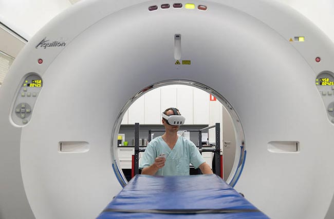
Equitom Equine Clinic, internationally recognized as the ‘clinic of last resort’, has reached a new milestone in veterinary medicine by becoming the first equine clinic in the world to integrate Virtual Reality (VR) into surgical and medical procedures. This advanced technology offers unprecedented opportunities to elevate the care and treatment of horses to an even higher level.
How are VR images for surgery created?
1. Medical Imaging as the foundation:
MRI and CT scans: The process begins with a CT scan (Computed Tomography) or an MRI scan (Magnetic Resonance Imaging) of the horse. These images provide a detailed 2D representation of the body’s internal structures, such as organs, bones, and tissues.
The collected images are then converted into highly detailed three-dimensional (3D) models that accurately depict the patient’s anatomy. Simultaneously, specific anatomical structures such as bones, muscles, blood vessels, etc., are separately identified and marked in 3D.
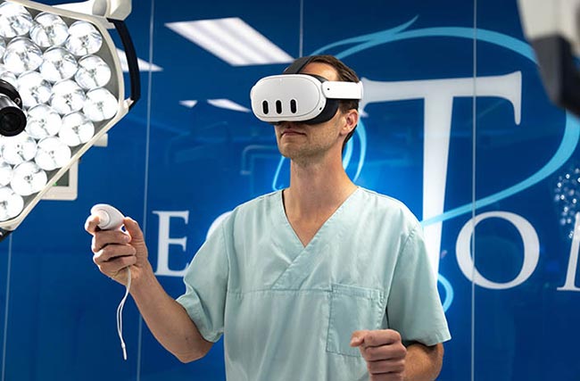
2. Virtual Reality Environment:
Integration into VR Platforms: The 3D models are then imported into a VR platform, where they are displayed in a virtual environment. This environment allows surgeons to view the patient’s anatomy from different angles, zoom in on specific areas, and even navigate through the virtual space as if they were inside the patient’s body.
Interaction: Surgeons can interact with the 3D models within the VR environment. For example, they can use a virtual scalpel to practice cutting, try out different surgical techniques, or simulate complex procedures without any risk to the patient... To read the complete article you need to be a subscriber
CLICK HERE TO SUBSCRIBE TO BREEDING NEWS
SUBSCRIBERS CAN READ THE COMPLETE ARTICLE BY LOGGING IN AND RETURNING TO THIS PAGE

