

By Dimitri Kadic MVM DES DACVS-LA DECVS
Photography: Courtesy Equitom
Guttural pouch mycosis is a condition unfortunately known only to few horse owners. The disease is gradually becoming more and more common in our regions and is usually fatal if not diagnosed in time.
It is a fungal infection that affects the horse’s guttural pouches, eventually resulting in swallowing problems and an unstoppable fatal nosebleed.
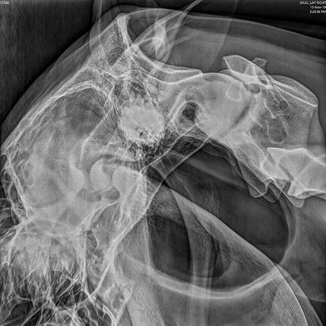
Radiographic image of the head and throat in a horse. The
radiolucent black area demonstrates the guttural pouches.
What are guttural pouches?
Every horse has a pair of guttural pouches located on either side of the throat area. They are sac-like bulges of the Eustachian tube. This tube forms a connection between the throat and the ear in the horse. The pouches are thus filled with air and are clearly observable radiographically as a radiolucent area (Photo 1). Their function is controversial to date, but what is certain is that some very important structures run along or through these pouches.
Blood vessels: Some of the main arteries of the head run along the wall of the guttural pouches. The main artery (arteria carotis communis) starts from the heart and carries oxygen-rich blood along the neck to the head and brain. At the level of the head, the main artery splits into several large vessels that are part of the wall of the guttural pouches (arteria carotis interna, arteria carotis externa and the arteria maxillaris) (Photo 2).
Nerves: There are 12 head nerves that start from the brain stem to innervate various structures. Some of these head nerves (IX, X, XII) cross the guttural pouches and can cause typical nerve symptoms in cases of guttural pouch mycosis. The glossopharyngeal nerve (IX) innervates the tongue muscles and throat. The vagus nerve (X) is an important nerve that innervates enormous muscles such as the vocal cords and larynx. The hypoglossal nerve (XII) innervates the tongue, among other anatomic structures (Photo 2).
In addition, some nerve centers of the autonomic nervous system (sympathetic ganglia) also lie in the vicinity of the guttural pouches.
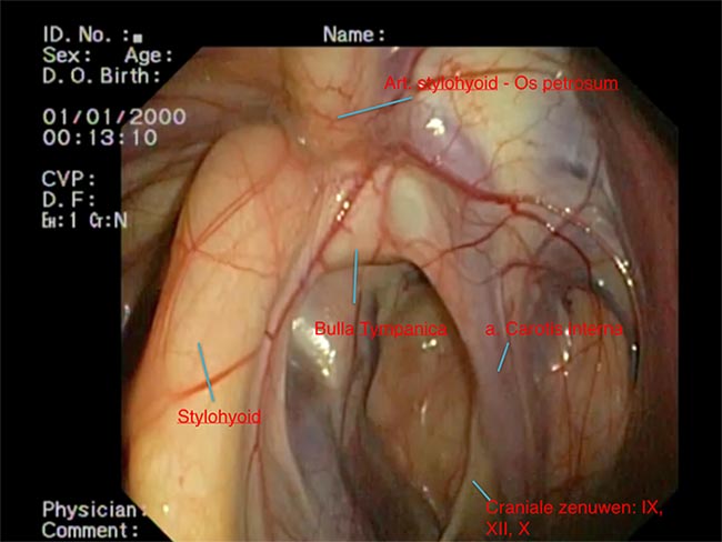
Endoscopic image of a normal guttural pouch. The hyoid bone (stylohyoid) lies centrally in the pouch and divides the it into two
compartments (lateral and medial). On the right side of the
stylohyoid we see the artery (arteria carotis interna) that is most colonized by the fungus. Next to this artery we see a fold in which some important nerves are located.
Fungal infection
As the name suggests, guttural pouch mycosis is a condition in which a fungal infection manifests itself at the level of these pouches. A typical fungus, Aspergillus fumigatus, is often isolated. Although this fungus is widespread throughout the world, it only settles in the pouches under certain conditions. Nobody knows yet why certain horses are affected, but it seems that certain climatic factors play a role. Increasing temperatures in our region due to global warming could explain the higher prevalence of this deadly disease in recent years.
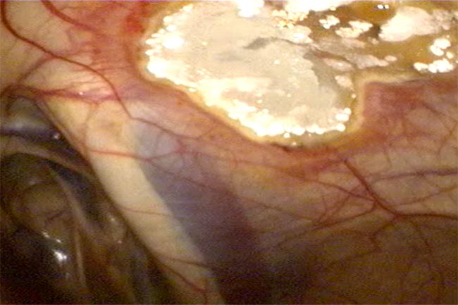
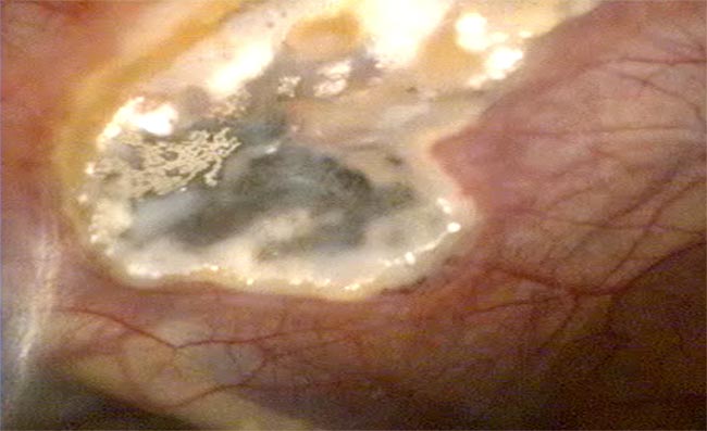
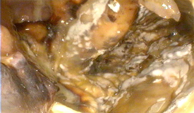
Aspergillus fungi, once established in the pouches, will seek a place where they can multiply optimally. Fungi like warm temperatures, and because there are large blood vessels in the guttural pouches, these are ideal nesting sites for the fungi. In addition, the arteries are also the ideal source of nutrients for the fungi. In most cases, the arteria carotis interna will be colonized by the Aspergillus (Photo 3 a and b, Photo 4).
Symptoms
Initially, the fungus will grow asymptomatically without being felt by the horse and without you as the horse owner noticing. This is precisely why this condition is so dangerous. Eventually, most horses get some nasal discharge. The problem is that numerous other rather harmless conditions such as sinusitis, pharyngitis, etc., can also be accompanied by nasal discharge as well so one is not immediately worried... To read the complete article you need to be a subscriber
CLICK HERE TO SUBSCRIBE TO BREEDING NEWS
SUBSCRIBERS CAN READ THE COMPLETE ARTICLE BY LOGGING IN AND RETURNING TO THIS PAGE

 awarded")
